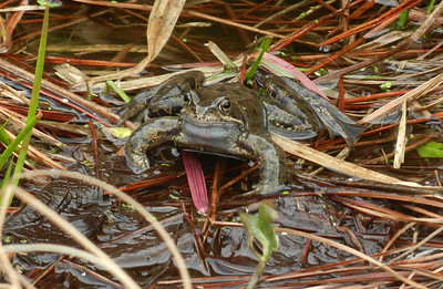
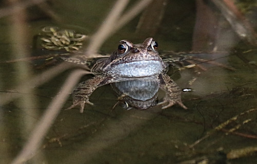
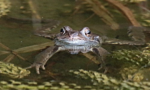
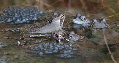
A second area of the pond is now filled with frog spawn. Most of the frogs dive under water when I go near but one was brave enough to stay put while I took a photo:
A frog he would a-wooing go,
Heigh ho! says Rowley,
A frog he would a-wooing go,
Whether his mother would let him or no.
With a rowley, powley, gammon, and spinach,
Heigh ho! says Anthony Rowley.
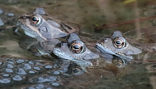
After hearing some croaking from the pond this morning I could see that the local frogs had definitely gone a-wooing.
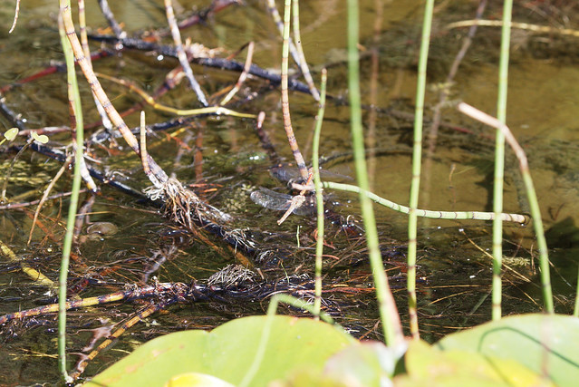
Pottering round the garden I suddenly noticed a pair of Common Darter Dragonflies. I have often seen them singly but this was the first time of seeing a pair together. Had to dash in to grab the Canon as the pocket Nikon would never have focussed on them:
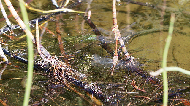
Not only that but the female kept dipping her 'tail' in the water:
Which I assume meant she was depositing eggs:
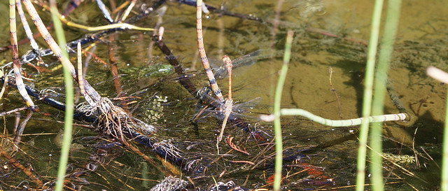
I was able to grab a few seconds of video before they moved on:
Here's hoping the pond is now a breeding ground for Dragonflies as well as Damselflies. Not forgetting the frogs and newts which also breed there.
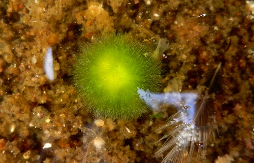
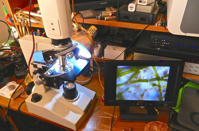
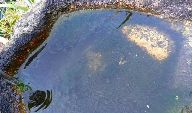
When I went to clean out the bird bath I noticed three green mounds in the water.
This was the largest at about 20mm (3/4 inch) across:
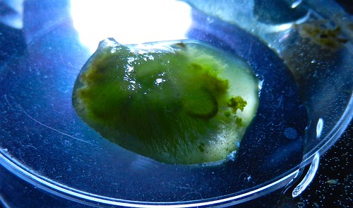
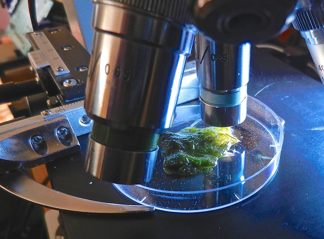
I decided it was time to have another microscope session to see what, if any, life forms were living in it. To that end I transferred the largest splodge to a plastic dish:
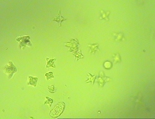
I had hoped to see a few bits of microscopic life but was blown away by their sheer numbers in such a small place as you will see later in the video:
A screen grab from the video. Each life form is less than a tenth of a mm in size.
I think the star shaped objects are Staurastrum:
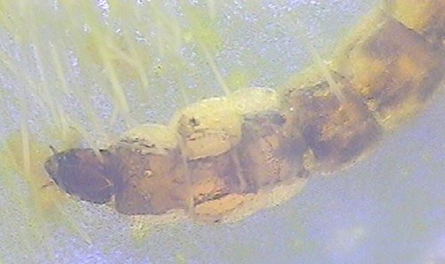
Another screen grab of a larger life form, about 10mm long and 1mm thick, possibly a midge larva. All the tiny pale specks are the things shown in the first screen grab:
On to the video:
Have a great weekend observing the wildlife around you, whatever its size.
Well, to be precise a Vorticella. A microscopic creature which looks a bit like an upside down bell. The top, the mouth opening, is surrounded by cilia which rapidly wave up and down in turn. This helps to suck minute food particles towards the opening. On the video they are moving so fast that at first glance I thought the top was rotating. From the bottom of the Vorticella is a stalk which it can use to attach itself and also coil it like a spring to rapidly move out of the way of danger.
By photographing the slide scale at the same size as the video view and making the background transparent it was possible to add the scale on the video. I tried for ages to get the background transparent in Affinity Photo but the result was poor so ended up going to my old copy of Elements which did it in three quick operations. Magic Wand to surround the black markings, hit delete to make the background see through. Save as .png. Job done!
From time to time I try different video cameras to find the best microscope / camera combination. All the cameras show some aberrations at low lighting levels. Can't quite make my mind up whether this is on the IR filter which they all have or some aspect of the sensor which only shows at low light levels.
This experiment was with a Siemens camera which is good quality but unfortunately shows a lot of aberrations. I have tried different high magnifications object lenses and that makes no difference.
This time I managed to obtain some closer views of the life in a bit of algae taken from the pond waterfall:
Once again I haven't managed to identify the various forms of life.
One of the things I had wanted to add to the microscope was a mechanical stage. This holds the slide or dish and has thumb screws for easy fine movement to position the specimen. They tend to be expensive but I spotted a universal fitting one on eBay and managed to secure it at the start price of £14.99. It is well constructed and a snip at the price. It has pins which fit in holes in the microscope stage to hold it firmly in place. Unfortunately although they were correctly space the holes in my microscope were smaller. Not dismayed I used a different locking screw and fitted an extra locking nut to hold it firmly without the pins. It is a delight to use and makes it so much easier to position things accurately under the lens, especially with high magnification lenses.
I had read about a technique for using bits of cotton on a slide to restrict the range of movement of live microscopic life. I wondered if the same would happen with some algae gathered from the pond. Off I went and grabbed a thumbnail size piece from the pool at the top of the pond waterfall:
Here are a few short extracts from the video clips I took this morning.
I haven't got round to identifying anything as yet.
Have a great weekend observing the wildlife around you, whatever its size.
A second session with the video camera on the microscope. This time with some water taken from the main pond:
During a search of the net I found a few pdf files to help me identify what I can see.
The first clip has a small round green diatom (bottom left) but I can't work out what the transparent creature is (centre screen).
The second clip shows several diatoms.
The final clip, my favourite, shows a rotifer (I hope).
All were observed using the same 100x objective with the video camera adding some more magnification as the view is larger than the sensor.
A few drops of water from my mini pond put under the microscope set up with the video camera:
To give an idea of size - the strap shaped object at about 43 seconds was approximately 2 mm long. That was the only thing I could see with the naked eye or even under a magnifying glass.
All the lenses are now working. The immersible ones are so close to the subject that they appear not to be working unless there is strong light from underneath. As there are no other lenses between the microscope and the video camera the magnification is not as great as it would be with a 'normal' set up. In some ways that is just as well as the little creatures soon disappear out of shot. The higher the magnification, the shallower the depth of field. As the creatures swim up and down they go in and out of focus.
What I really want is a cross table for the microscope. That allows fine movement to position the specimen which is needed at high magnification. Moving things by hand is very much hit and miss as a mm or two of movement can make the difference between seeing and not seeing a particular part.